Home
Orthodontics - Orthognathic surgery
- Introduction
- Surgical patient's guide
- My story
- OCTOBER 20, 2006 - The installation of my first braces
- DECEMBER 8, 2006 - The installation of my lower braces
- DECEMBER 13, 2007 - The removal of my wisdom teeth
- MARCH 15, 2008 - My surgery (2 days before my first surgery)
- MARCH 17, 2008 - News about Marie-Hélène (email sent by my husband)
- MARCH 19, 2008 - Update (email sent by my husband)
- MARCH 19, 2008 - Update (Take 2) (email sent by my husband)
- MARCH 21, 2008 - News from the surgical patient!
- MARCH 25, 2008 - Other news and first new pictures
- APRIL 1, 2008 - Nothing but good news
- APRIL 8, 2008 - Latest news
- APRIL 14, 2008 - Not so good news
- APRIL 15, 2008 - RE: ** UPDATE ** Not so good news
- APRIL 15, 2008 - News from the second surgery (email sent by my husband)
- APRIL 18, 2008 - News from the one that is slowly recuperating from her second surgery
- APRIL 22, 2008 - News... again!
- MAY 1, 2008 - After 6 weeks of appointments...
- JUNE 6, 2008 - On the right track
- AUGUST 27, 2008 - News!
- NOVEMBER 17, 2008 - Another ordeal
- DECEMBER 17, 2008 - Good news before Christmas
- FEBRUARY 10, 2009 - The ugly duckling turned into a swan
- FEBRUARY 9, 2012 - My teeth are acting up again!
- FEBRUARY 13, 2012 - Appointment with my surgeon for my jaws
- MARCH 20, 2012 - I will probably get out of it without braces!
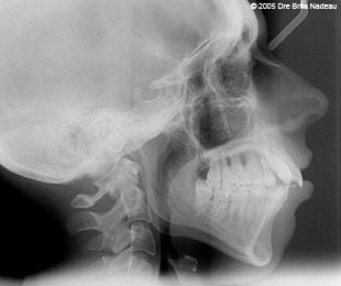
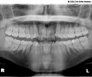
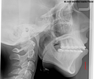
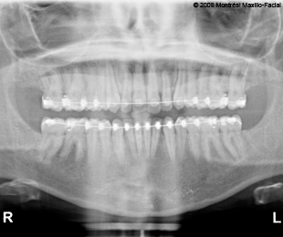
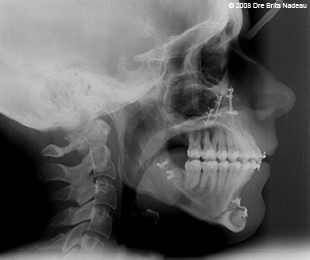
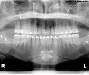
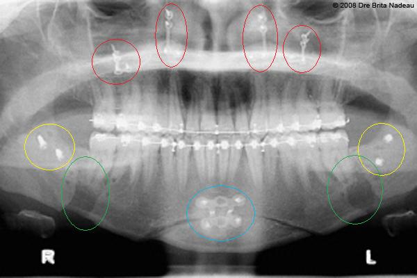
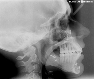
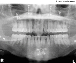
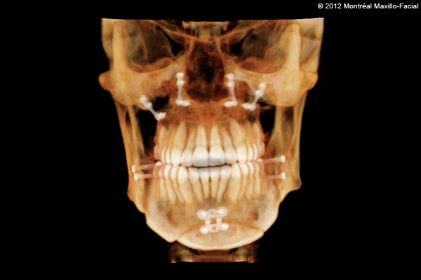
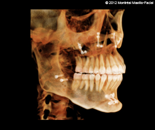
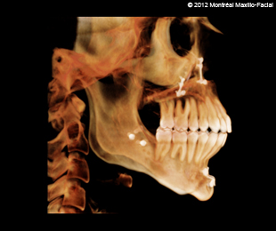
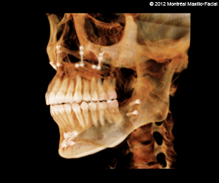
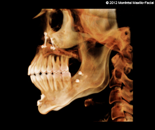
- Pictures & X-rays
Bon Jovi
- Introduction
- My show reviews
- DECEMBER 10, 1993 - Montreal, Quebec, Canada
- AUGUST 3, 1995 - Montreal, Quebec, Canada
- NOVEMBER 28, 2000 - Montreal, Quebec, Canada
- MAY 19, 2001 - Quebec City, Quebec, Canada
- JULY 19, 2001 - Montreal, Quebec, Canada
- FEBRUARY 21, 2003 - Montreal, Quebec, Canada
- DECEMBER 14 & 15, 2005 - Montreal, Quebec, Canada
- JULY 13, 2006 - Montreal, Quebec, Canada
- NOVEMBER 14 & 15, 2007 - Montreal, Quebec, Canada
- MARCH 19 & 20, 2010 - Montreal, Quebec, Canada
- MAY 27, 2010 - East Rutherford, New Jersey, USA
- JULY 24, 2010 - Foxborough, Massachusetts, USA
- FEBRUARY 18 & 19, 2011 - Montreal, Quebec, Canada
- MAY 4, 2011 - Montreal, Quebec, Canada
- JULY 9, 2012 - Quebec City, Quebec, Canada
- FEBRUARY 13 & 14, 2013 - Montreal, Quebec, Canada
- APRIL 20, 2013 - Las Vegas, Nevada, USA
- NOVEMBER 1 & 2, 2013 - Toronto, Ontario, Canada
- NOVEMBER 8, 2013 - Montreal, Quebec, Canada
- APRIL 7, 2018 - Newark, New Jersey, USA
- MAY 17 & 18, 2018 - Montreal, Quebec, Canada
- My Bon Jovi pictures
- DECEMBER 14, 2005 - Montreal, Quebec, Canada
- DECEMBER 15, 2005 - Montreal, Quebec, Canada
- JULY 13, 2006 - Montreal, Quebec, Canada
- NOVEMBER 14, 2007 - Montreal, Quebec, Canada
- NOVEMBER 15, 2007 - Montreal, Quebec, Canada
- MARCH 19, 2010 - Montreal, Quebec, Canada
- MARCH 20, 2010 - Montreal, Quebec, Canada
- MAY 27, 2010 - East Rutherford, New Jersey, USA
- JULY 24, 2010 - Foxborough, Massachusetts, USA
- FEBRUARY 18, 2011 - Montreal, Quebec, Canada
- FEBRUARY 19, 2011 - Montreal, Quebec, Canada
- MAY 4, 2011 - Montreal, Quebec, Canada
- JULY 9, 2012 - Quebec City, Quebec, Canada
- FEBRUARY 13, 2013 - Montreal, Quebec, Canada
- FEBRUARY 14, 2013 - Montreal, Quebec, Canada
- APRIL 20, 2013 - Las Vegas, Nevada, USA
- NOVEMBER 1, 2013 - Toronto, Ontario, Canada
- NOVEMBER 2, 2013 - Toronto, Ontario, Canada
- NOVEMBER 8, 2013 - Montreal, Quebec, Canada
- APRIL 7, 2018 - Newark, New Jersey, USA
- MAY 17, 2018 - Montreal, Quebec, Canada
- MAY 18, 2018 - Montreal, Quebec, Canada
- My Bon Jovi videos
- DECEMBER 14, 2005 - Montreal, Quebec, Canada
- DECEMBER 15, 2005 - Montreal, Quebec, Canada
- JULY 13, 2006 - Montreal, Quebec, Canada
- NOVEMBER 14, 2007 - Montreal, Quebec, Canada
- NOVEMBER 15, 2007 - Montreal, Quebec, Canada
- MARCH 19, 2010 - Montreal, Quebec, Canada
- MARCH 20, 2010 - Montreal, Quebec, Canada
- MAY 27, 2010 - East Rutherford, New Jersey, USA
- JULY 24, 2010 - Foxborough, Massachusetts, USA
- FEBRUARY 18, 2011 - Montreal, Quebec, Canada
- FEBRUARY 19, 2011 - Montreal, Quebec, Canada
- MAY 4, 2011 - Montreal, Quebec, Canada
- JULY 9, 2012 - Quebec City, Quebec, Canada
- FEBRUARY 13, 2013 - Montreal, Quebec, Canada
- FEBRUARY 14, 2013 - Montreal, Quebec, Canada
- APRIL 20, 2013 - Las Vegas, Nevada, USA
- NOVEMBER 1, 2013 - Toronto, Ontario, Canada
- NOVEMBER 2, 2013 - Toronto, Ontario, Canada
- NOVEMBER 8, 2013 - Montreal, Quebec, Canada
- APRIL 7, 2018 - Newark, New Jersey, USA
- MAY 17, 2018 - Montreal, Quebec, Canada
- MAY 18, 2018 - Montreal, Quebec, Canada
- My texts and poetry
- That Power Called Love (1997)
- For My 5 Years of Devotion (1998)
- 6 Years (1999)
- I Love You Because (2000)
- 10 Years - One Decade (2003)
- Already 10 Years of Love (2003)
- Deception of a Fan (2003)
- Living In a Dream (2003)
- You and I (2003)
- Time Goes By Fast... When You're in Love (2004)
- For Half My Life... (2005)
- Have a Nice Day Farewell (2006)
- 15 Years Ago ... on February 21, 1993... (2008)
- My Love Celebrates Its Sweet 16 (2009)
- What You Mean to Me (2011)
Literature
About me


